Bacterial diseases of tomato: bacterial spot, bacterial speck and bacterial canker
Learn about the common bacterial diseases of tomatoes in Ontario.
ISSN 1198-712X. Published 2005.
Introduction
Three bacterial diseases are common in Ontario tomato fields:
- bacterial spot, caused by Xanthomonas campestris pv. Vesicatoria
- bacterial speck, caused by Pseudomonas syringae pv. tomato
- bacterial canker, caused by Clavibacter michiganensis subsp. michiganensis.
In Ontario, bacterial disease is present at some level every season, though not always at destructive levels. However, when conditions are optimal for bacterial disease, losses in marketable yield can be up to 60% in some fields.
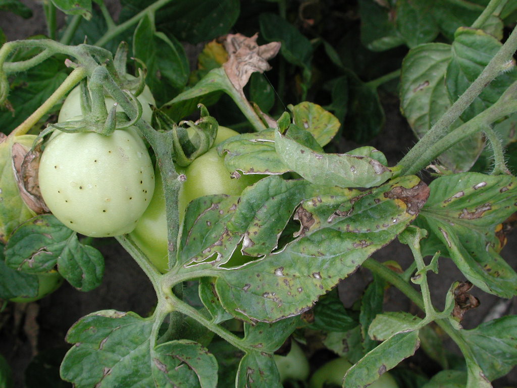
Damage from these diseases may range from a light spotting of the foliage to almost complete defoliation of the plant, with corresponding impacts on photosynthesis and production potential. When present, fruit lesions disfigure and reduce the marketability of both fresh-market and processing fruit (especially in whole-pack or diced product) and interfere with peeling. Defoliation exposes the fruit, resulting in sunscald and poor colour. Secondary rots can also develop.
Both fresh-market and processing growers may incur higher sorting costs due to fruit lesions. Processing growers also face the risk of increased tare penalties and the possibility of not meeting their contracted tonnage. Depending on the product being produced, bacterial disease may result in lower solids, increased costs, slower factory operations and reduced peeled recovery for the processors. Processors also face the risk of falling short of their packing goals.
Management of tomato bacterial diseases must focus on prevention and must start well before transplanting. Seed suppliers, transplant growers, field growers, processors, researchers, extension specialists and crop advisors all have a part to play.
The pathogens
Bacterial pathogens need moisture to multiply. Wet conditions in the plant canopy due to rain, fog, dew, high humidity or irrigation give the bacteria a suitable environment for growth. Each pathogen has a particular temperature range, in which it is at its peak rate of growth and infection (see Table 1). The pathogens multiply much more slowly outside this optimum range.
| Disease | Causal organism | Optimum temperature |
|---|---|---|
| bacterial spot | Xanthomonas campestris pv. vesicatoria | 24°C to 30°C |
| bacterial speck | Pseudomonas syringae pv. tomato | 18°C to 24°C |
| bacterial canker | Clavibacter michiganensis subsp. michiganensis | 24°C to 32°C |
Bacterial spot
Symptoms
The bacterial spot pathogen may produce lesions on all aboveground parts of the plant - leaves, stems, flowers and fruit. It is difficult to reliably distinguish bacterial spot from bacterial speck based on visual symptoms, especially in the early stages.
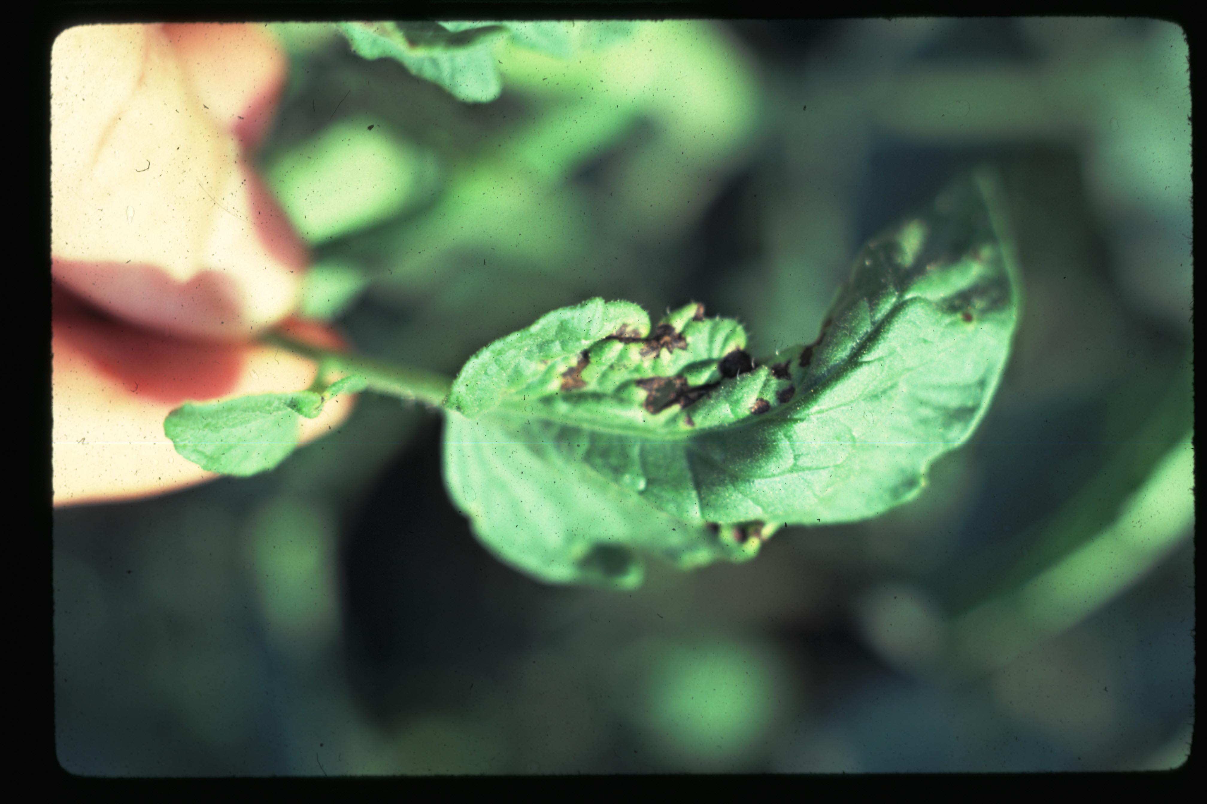
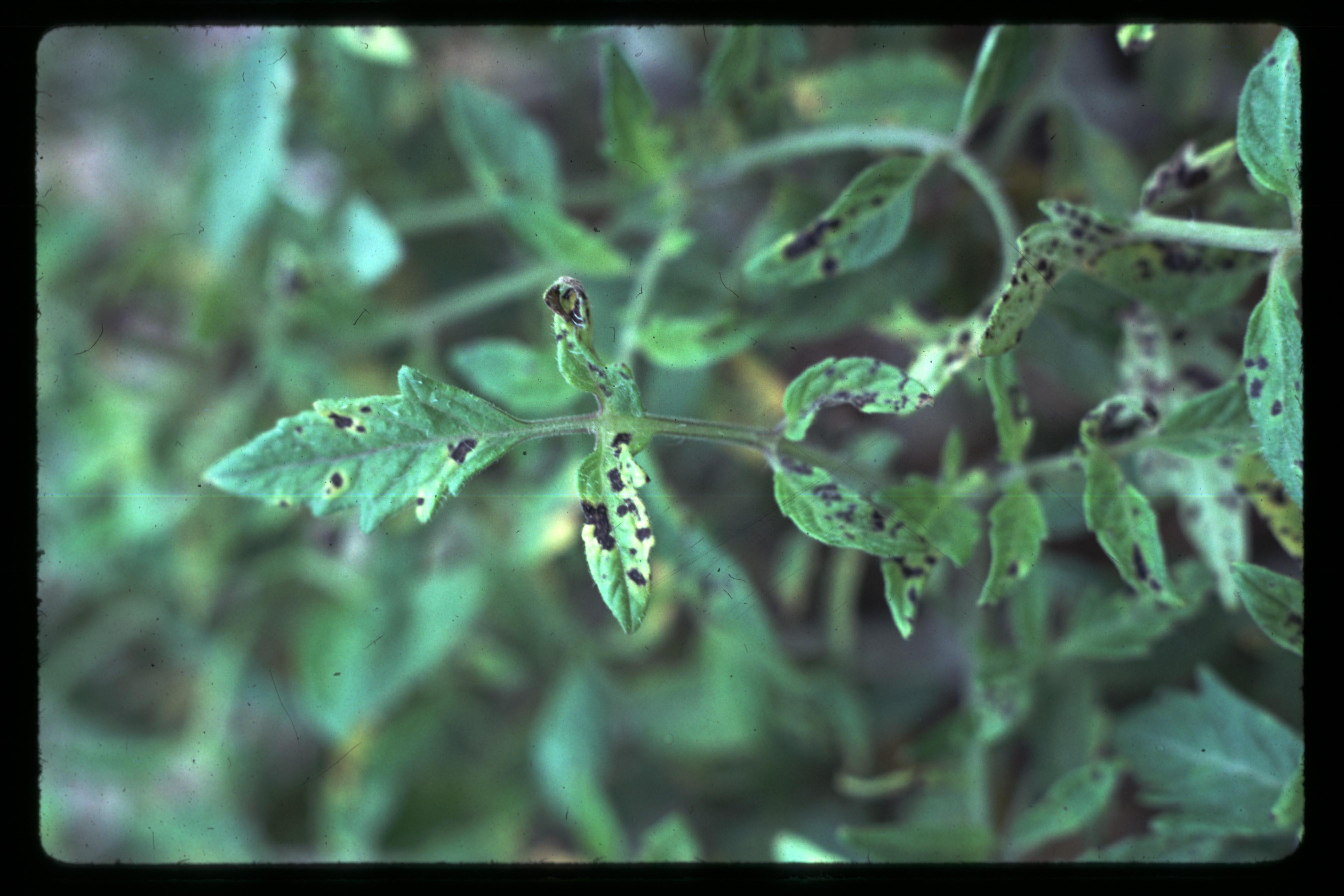
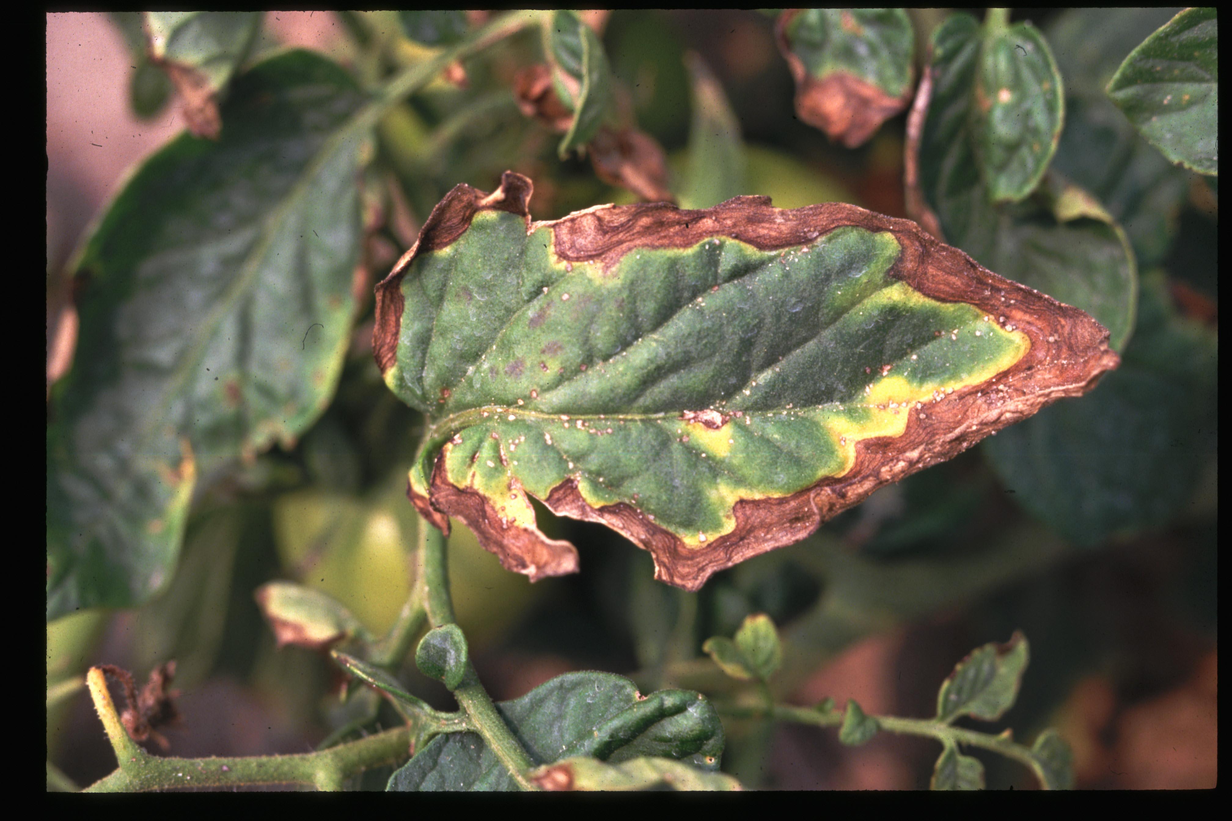
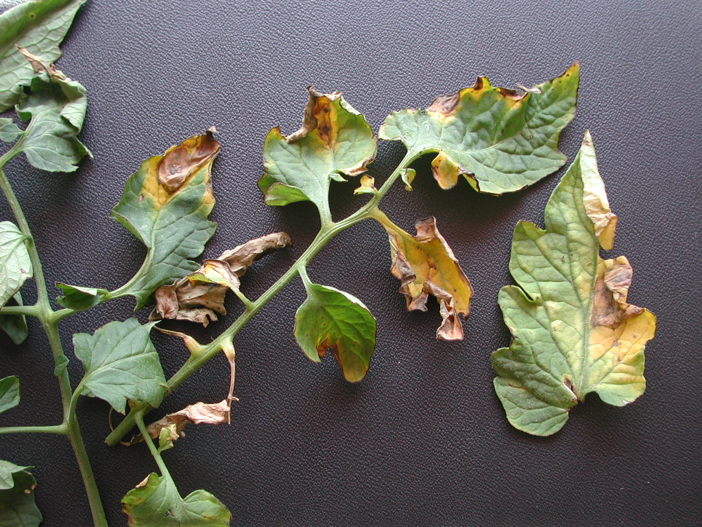
Initial leaf symptoms are small, circular-to-irregular, dark lesions, which may be surrounded by a yellow halo. The lesions tend to concentrate on the leaf edges and tip and may increase in size to a diameter of 3-5 mm. Infected leaves may develop a scorched appearance. When spots are numerous, foliage turns yellow and eventually dies, leading to defoliation of the lower portion of the plant.
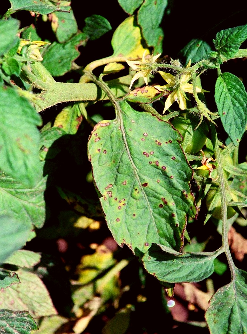
Lesions on pedicels may cause flower abortion, resulting in lost yield and split sets of fruit.
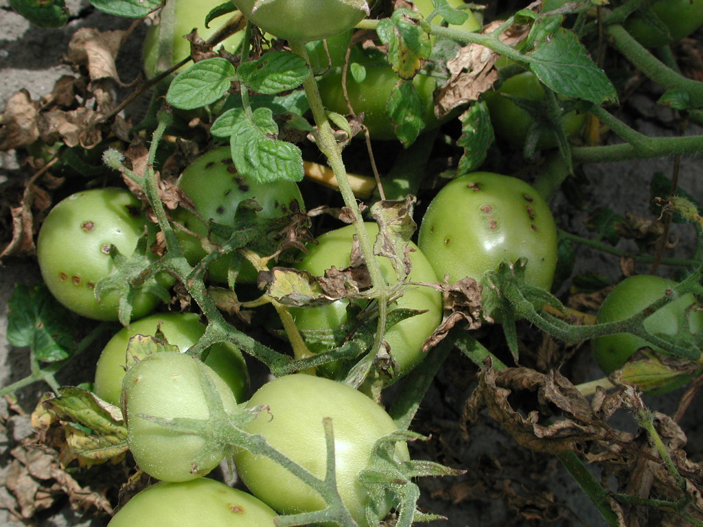
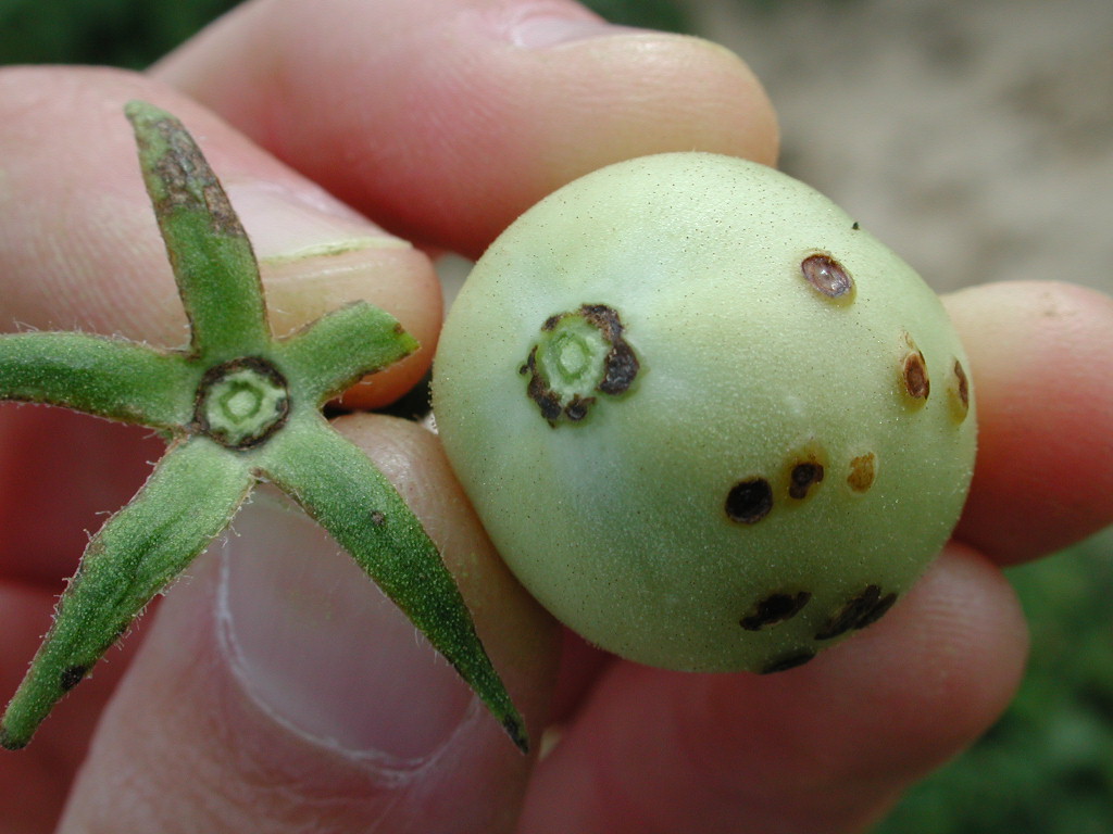
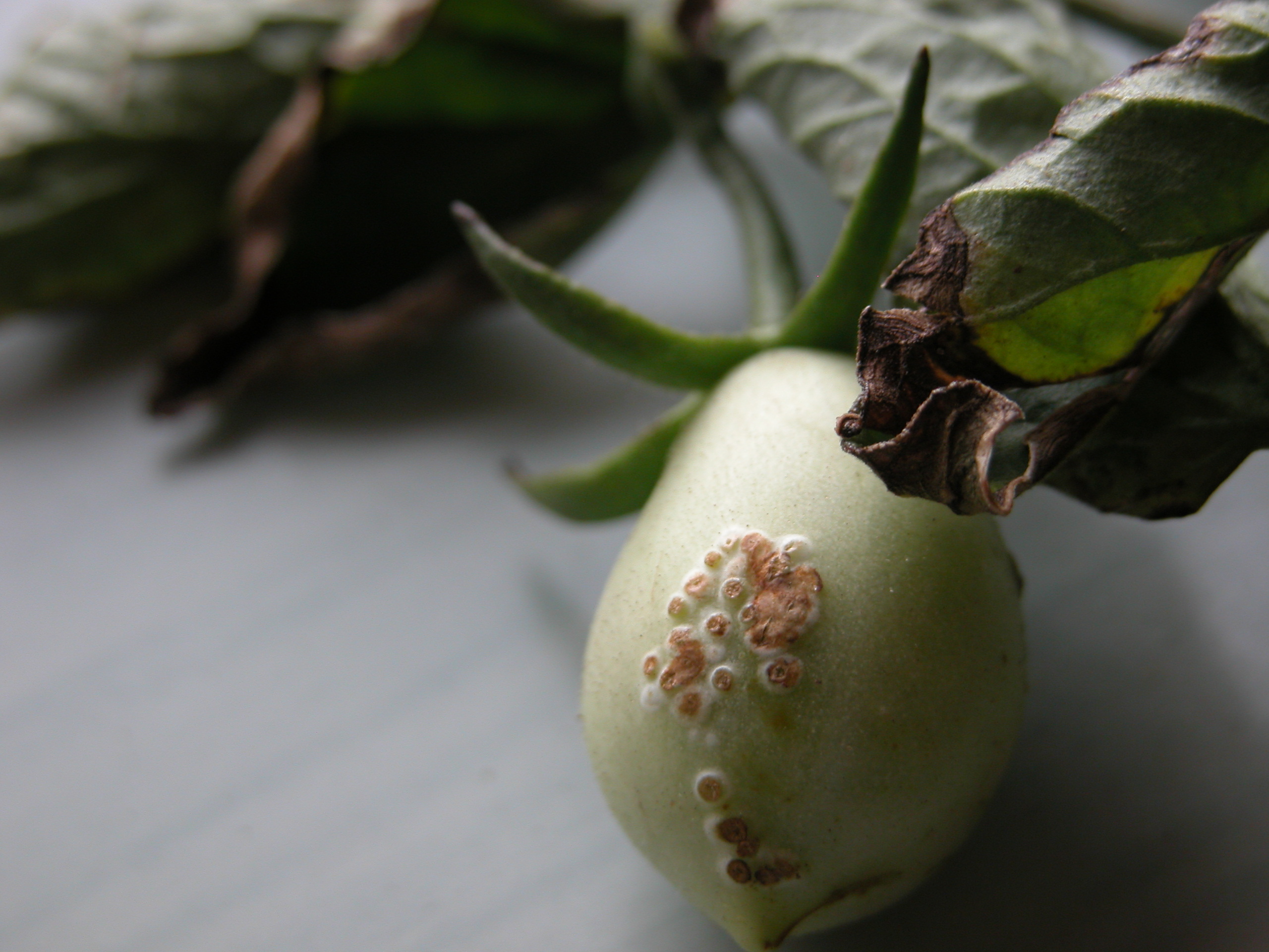
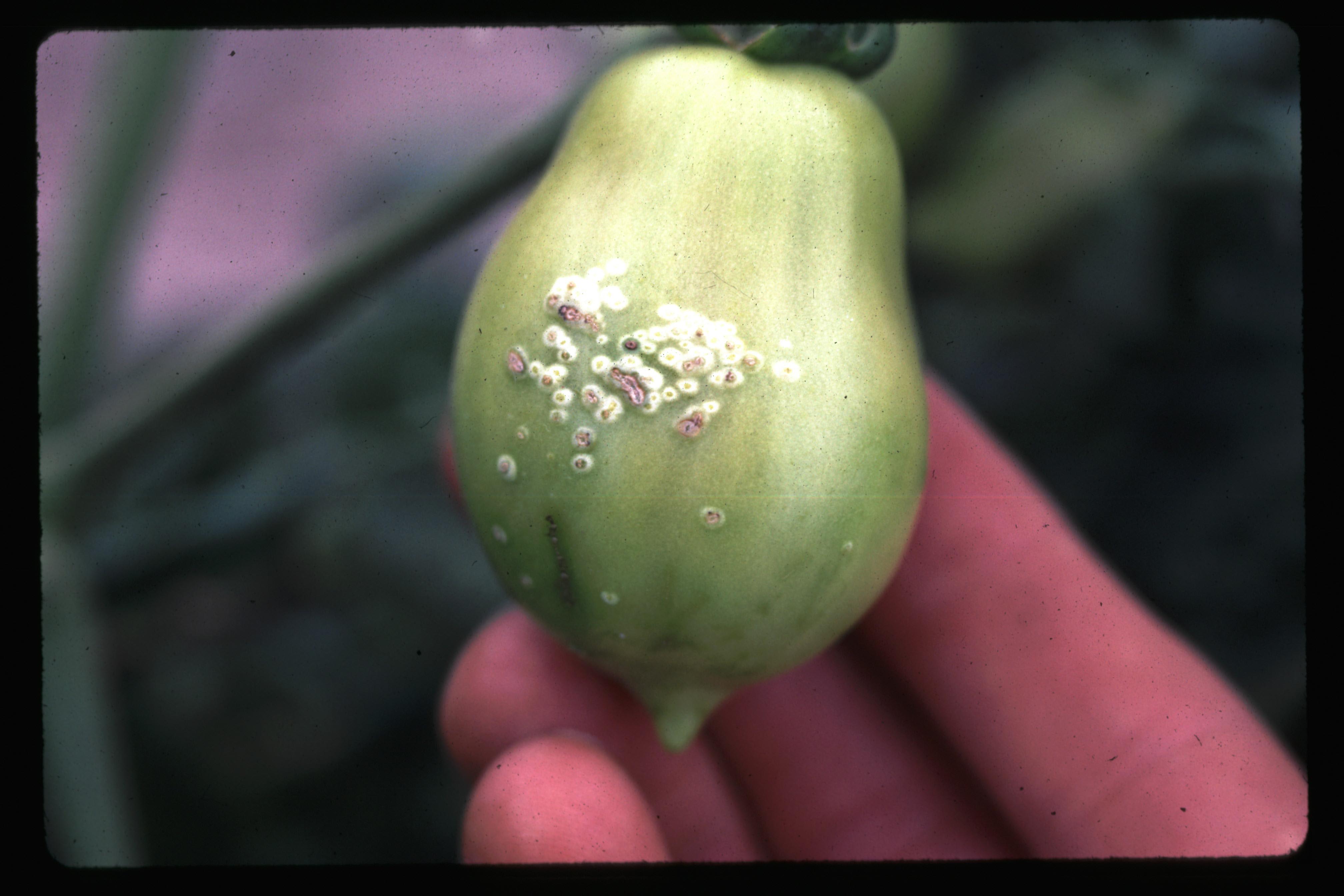
Fruit lesions are initiated only on green fruit, most likely because infection occurs through fruit hairs, which are present only on immature fruit. On fruit, the first symptoms are small, dark brown-to-black, raised spots. The lesions also may have a white halo, similar to the bird's-eye spotting seen with bacterial canker. As the fruit ages, the white halos disappear. In contrast, bacterial canker fruit lesions retain their white halo. Bacterial spot lesions may increase in size to 4-6 mm in diameter and become brown, greasy-looking and sometimes scabby.
Inoculum and spread
The major sources of infection for these bacteria are thought to be seed and infected crop debris. Like the bacterial speck pathogen, they also may be present on volunteer tomato plants and on the surfaces of contaminated equipment (farm machinery, racks, greenhouse structures, tools). The bacteria are spread primarily by splashing water and wind-driven rain or mists produced during storms. In the field, spread by equipment or workers is probably of lesser importance than it is in the greenhouse, unless wounds are being opened up at the same time, as in a pruning operation or when plants are injured by a cultivator.
Bacteria enter the plant through natural openings (stomates and hydathodes) or wounds caused by wind-driven soil, insects or mechanical damage (handling, wind whipping, high pressure sprayers).
Causal organism
The bacteria that cause bacterial spot are called xanthomonads. Recent taxonomic studies have indicated that these bacteria belong to one of four groups: A, B, C and D. The original (and still valid), taxonomic name given to these bacteria was Xanthomonas campestris pv. vesicatoria, but recent work has indicated that each group may represent a separate species. Group D has become the predominant form in Ontario. This is of concern, as recent research has shown it to be a particularly aggressive form, which can overwinter under southern Ontario conditions. The bacterial spot-causing xanthomonads also have been classified into races, some of which infect only tomato or pepper, and some of which can infect both crops.
The genetic variability of the bacterial spot-causing xanthomonads makes it difficult for plant breeders to develop stable resistance in tomato varieties and for pathologists to develop control measures. If genetic resistance or chemical controls are only effective on one strain, the pathogen population will simply shift to the more tolerant strains.
Bacterial speck
Symptoms
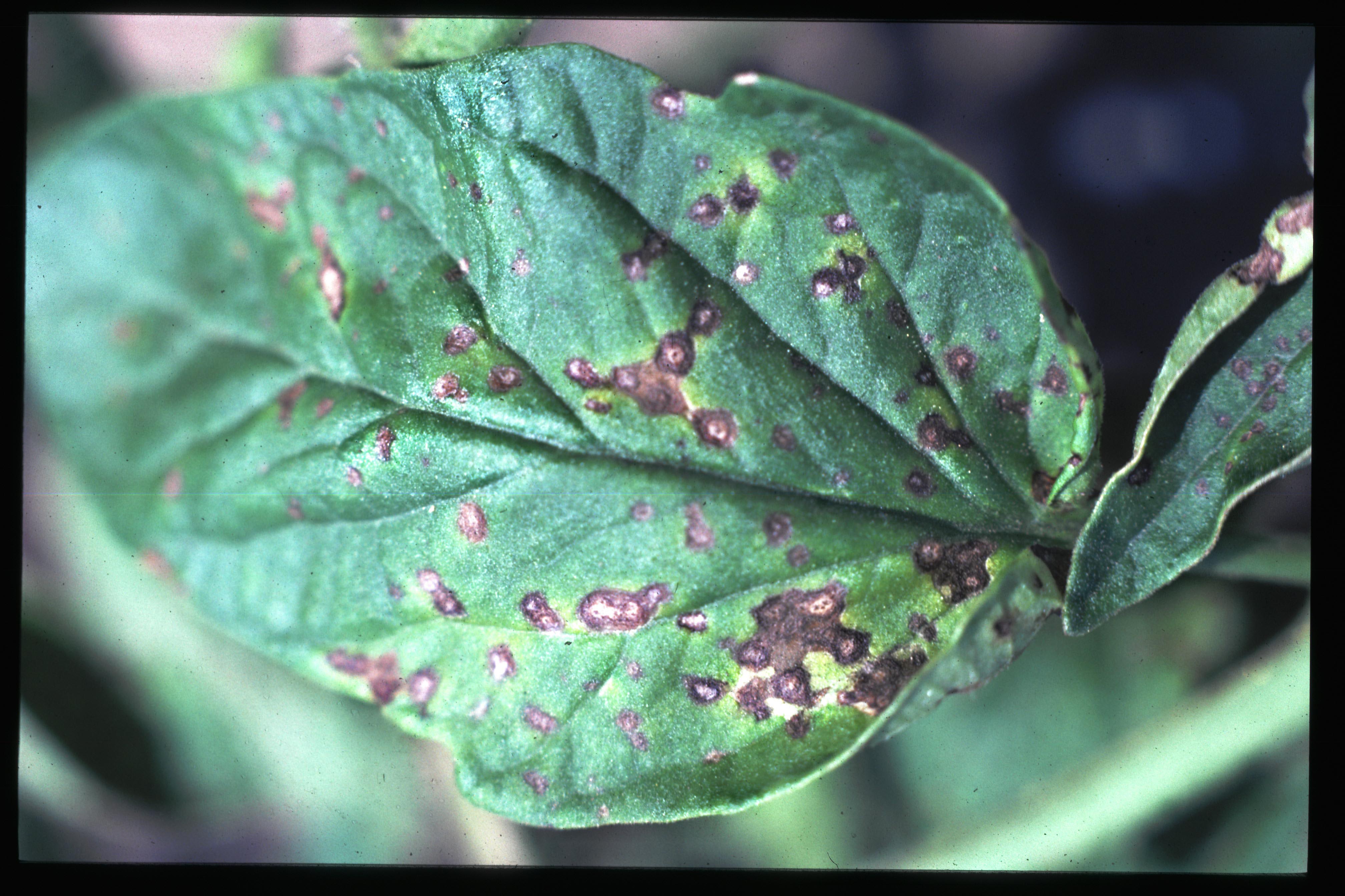
Bacterial speck lesions may occur anywhere on the foliage, stems or fruit. Symptoms are very difficult to visually distinguish from bacterial spot and can be confused with young, early blight lesions. On leaves, symptoms appear as black specks, usually no more than 2 mm in diameter, which are usually surrounded by a yellow halo. Speck lesions sometimes cause distortion of the leaf, as the infection restricts the expansion of leaf tissue. Lesions are often concentrated near leaf edges, and in some cases, leaf margin burn resembling bacterial canker may occur. When numerous, lesions may coalesce, and entire leaflets may die. Severely infected seedlings may become stunted.
Only green fruit less than 3 cm in diameter is susceptible to infection by the bacterial speck pathogen. Small (less than 1-3 mm), slightly raised black specks develop and are often surrounded by a narrow green to yellow halo. Lesions are usually superficial and can be scraped off with a fingernail. Red fruit are not susceptible to infection, likely due to a lack of entry points for bacteria; fruit hairs, which may break and allow bacteria to enter, are only present on young fruit. On fruit previously infected, black lesions remain after ripening.
Inoculum and spread
The sources of bacterial speck inoculum and the methods of spread are the same as those for bacterial spot. Studies have shown that the speck organism can survive in the crevices and cavities of the tomato seed coat for up to 20 years.
Causal organism
Bacterial speck is caused by Pseudomonas syringae pv. tomato. Two races are present in Ontario, race 0 and race 1. The Pto gene, discovered by Ontario researchers, confers resistance to race 0.
This bacterium produces a number of compounds that help it infect and obtain nutrients from the tomato plant. One of these compounds is the plant-specific toxin coronatine, which is responsible for the yellow halo surrounding leaf lesions and the stunting of young seedlings. The majority of bacterial speck strains that have been isolated from Ontario tomato fields are either resistant to or tolerant of copper-based bactericides.
Bacterial canker
Symptoms
Bacterial canker, which may occur in tomato as a primary (systemic) or secondary (foliar) infection, shows a wide range of symptoms.
Primary infections originate from infected seed or from invasion of the vascular tissue of young seedlings. Symptoms, which may not show up until several weeks after infection, initially appear as wilting and downward turning of the lower leaves. The wilting generally progresses upwards, unless the site of infection is in the upper part of the plant. Wilting is often seen only on one side of the leaf or one side of the plant. Plants may collapse and die, especially if infected at a very early stage. Generally, plants survive but are stunted, showing some or all of the symptoms described here, depending on their environment and stage of growth.
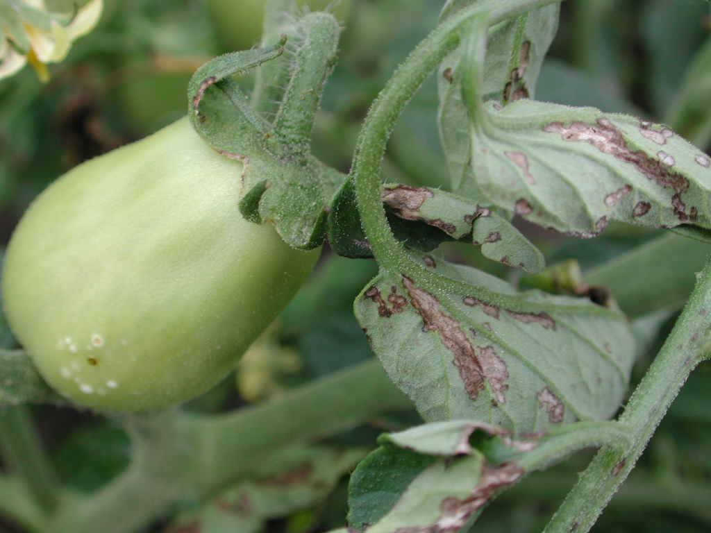
Tomato foliage infected with the canker organism has distinctive black leaf edges with no spotting on the interior of the leaves. Sometimes a thin yellow border is present between the dead leaf margins and healthy tissue.
If an infected stem is cut lengthwise, a light brown discolouration may be present in the vascular tissue, most noticeable at nodes and just above the soil line. As the disease progresses, this turns reddish-brown. Light coloured streaks are often visible on the outside of the stem. These may later darken and break open into cankers. With severe infections, a yellow ooze may exude from a cut stem when it is squeezed.
Fruit may develop relatively small spots with light brown centres, generally surrounded by a greasy white halo (3-6 mm in diameter). These are known as bird's-eye spots. With bacterial canker lesions, this white halo generally remains as the fruit ripens, while in the case of bacterial spot, it disappears with time. Bacterial canker may also cause a darkening of the vascular tissues within the fruit. The fruit may show a black peppering at the vascular bundles under the calyx scar. Canker bacteria can grow in the vascular bundles within the fruit, all the way to the seed. This can result in visible yellowish strands from the stem to the seeds and internal infections in the seed.
With a secondary foliar infection, leaves develop brown-black margins with a thin, yellow (chlorotic) band. Leaflet edges may curl upwards. Fruit may show bird's-eye spotting, as in a systemic infection. Secondary infections (no vascular system involvement) often have minimal impact on the crop, especially when initiated later in the season.
Inoculum and spread
Infected seed is probably the major source for primary (systemic) infections. The bacteria can be present on the surface of the seed as well as within the innermost layer of the seed coat. This makes the canker organism harder to eradicate with seed treatments than the spot and speck pathogens.
The organism can also be introduced from infected crop debris, weed hosts or volunteer tomatoes, and contaminated equipment. Studies in the U.S. northern Midwest have shown that it can overwinter on infected debris. However, crop rotation and tillage before planting should reduce the risk of infection from this source.
The canker bacteria enter the plant through natural openings and wounds, including root wounds. Pruning or transplant clipping operations can introduce the bacteria directly into the vascular system, resulting in the more serious systemic infections.
Infections spread through splashing water, wind-driven rain and the fine water droplets or aerosols produced during storms. In the field, bacteria transfer by machinery or workers is probably not as significant as in the transplant greenhouse where plant density is high and growth conditions for the bacterium are optimal.
Causal organism
Bacterial canker is caused by Clavibacter michiganensis subsp. michiganensis.
Diseases and disorders with similar symptoms
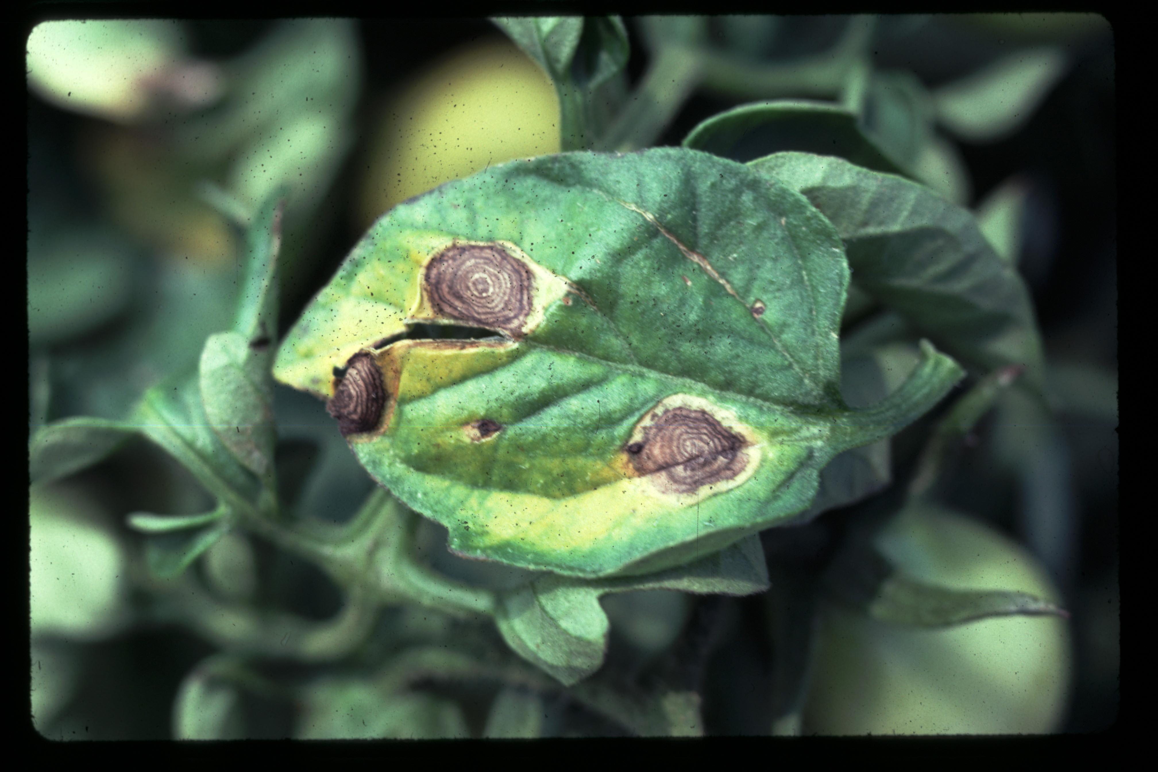
Early blight and septoria leaf spot are two common fungal diseases that cause spots on tomato foliage. Young early blight lesions can resemble bacterial lesions and often have a yellow halo. Look for the dark concentric rings that indicate early blight. Early blight lesions enlarge and become angular over time. Septoria lesions can be distinguished due to their light tan centres containing tiny black dots (pycnidia). These can be seen clearly with a hand lens. Yellowing of the foliage is rarely present with septoria leaf spot until lesions become numerous. Neither fungal disease produces the small black fruit lesions typical of speck or spot, or the bird's-eye spotting of canker.
Ozone injury may also cause spotting on tomato foliage, along with yellowing, purpling, glazing or curling of leaves. Recently matured leaves are most susceptible, and injury may also be present on nearby sensitive species. On fruit, hail damage and insect stings can resemble bacterial lesions.
The wilting symptoms of a systemic bacterial canker infection may be confused with verticillium wilt, which also may cause wilting on one side of the plant or leaf and browning of the vascular system near the soil line. Verticillium wilt typically causes significant yellowing of the foliage and V-shaped lesions extending out to the leaf tips. Another wilt disease, bacterial wilt, causes a more extensive discolouration of the vascular and stem tissue, which may extend well below the soil line. Bacterial wilt does not overwinter in Ontario, and so would only be found on transplants from southern US growing regions. (One race of bacterial wilt (race 3 biovar 2) may be able to overwinter in northern regions, but this form has not become established in the US or Canada. This race is a regulated and quarantined pest.) Lightning injury and walnut wilt (of plants growing near black walnut trees) are other common causes of wilt symptoms.
Diagnosis
Sampling
For plant disease diagnosis, choose representative plants showing early symptoms. Submit as much of the plant as is practical, or several plants showing a range of symptoms.
Wrap plants in newspaper and put in a plastic bag. Tie the root system off in a separate plastic bag to avoid drying out and contamination of the leaves by soil. Do not add moisture, as this encourages decay in transit. Cushion specimens and pack in a sturdy box to avoid damage during shipping. Protect specimens from excessive heat or freezing and deliver to the diagnostic lab as soon as possible by first class mail or courier at the beginning of the week.
Diagnostic services
Ontario Ministry of Agriculture, Food and Rural Affairs (OMAFRA) Publication 838, Vegetable Crop Protection Guide, provides information on pest diagnostic services in Ontario.
Management
General strategies
To develop an effective disease management strategy, it is important to know how the bacterial pathogens differ from the fungal pathogens. Fungal pathogens multiply by spores, which are carried by wind or other means to new host plants, where they germinate and grow directly into the plant tissue. Bacterial pathogens are spread primarily by water and can travel long distances in wind-driven rain. Sprinkler irrigation systems also provide a means of disseminating bacteria. Once they arrive on the plant surface, bacteria must find a wound or natural opening to enter the plant and start the disease process. Bacteria multiply much more rapidly than fungi; under optimal conditions producing a new generation every 90 min.
Although bacteria are relatively easy to kill with disinfectants when they are on smooth, non-living surfaces, they are difficult to eradicate when they are on plants. Disinfectants typically cannot be used on a crop, because they may damage plant tissue. Fixed copper must come in direct contact with the bacteria to be effective. Bacteria inside the leaf (in stomates, hydathodes or wounds) and even in crevices on the leaf surface are protected and continue to multiply. In contrast, with fungal disease, preventing spore germination or mycelial growth with fungicide controls infection and spread.
Management strategies for tomato bacterial disease control must take a multifaceted approach. Since it is difficult to eliminate bacteria from the crop once they are established, the first step is to try to exclude the bacteria in the first place. We do not presently have a procedure that can cost-effectively screen enough seedlings to make sure we are free of the pathogens, so we must also employ a preventative program early in the crop's development.
Experience has shown that if a bacterial disease outbreak can be delayed until after the main fruit set, the crop will be minimally affected. Once the plant has reached a full canopy, a low level of bacterial disease on the foliage can be tolerated. Fruit lesions, which have a major impact on marketable yield, can only be initiated on young green fruit, so control measures used prior to fruiting are most beneficial.
Plants under stress are more susceptible to disease than plants growing under ideal conditions. Some environmental stresses are out of the grower's control, but management practices can reduce many stress factors, including those associated with dry weather, excess water, compacted or poorly structured soils, weeds, insects, other diseases, and field operations. Bacterial disease often seems to affect early cultivars most severely. The cool temperatures and stressful conditions frequently experienced by early plantings are likely a contributing factor.
The specific management recommendations below are based on an understanding of the biology of the pathogens and the response of the crop, as described above.
Cultural
Seed
Ideally, seed from disease-free fields would be used for all of our tomato crops. In reality, we can never be certain that our seed is completely clean, so additional measures are taken to reduce the risk of disease.
Seed extraction alone, whether through fermentation or acid methods, is not a reliable method of eliminating bacteria on the seed. Extraction should be followed by a hot water, acid or chlorine disinfection. All seed lots should be disinfected by one of these methods.
The hot water method is the least desirable, as temperature and duration are limited by the need to keep seeds viable. On the other hand, hot water treatment can be done on-farm, with equipment that is relatively easy to obtain and is better than no treatment at all. Instructions must be followed carefully to minimize damage to the seed. Even so, significant losses of germination could occur in some seed lots.
Acid and chlorine disinfection are very difficult to do on-farm. Seed can be damaged if protocols are not followed exactly. Generally, growers must trust their seed supplier to perform the seed disinfection effectively.
The acid and chlorine treatments are very effective on spot and speck if done correctly. Some canker bacteria, however, may survive, as this organism has been found inside the seed coat.
Effective seed disinfection is critical to bacterial disease management in tomatoes. Talk to your seed supplier about the disinfection procedure used on your seed lots. Your supplier should be able to provide the following information:
- What disinfection method was used? Hot water, acid and chlorine treatments are discussed above. Researchers are studying the use of other disinfectants and alternative methods including microwave, sonication and hydrostatic pressure treatments.
- How do you ensure that all the seeds receive adequate treatment? Presoaking is necessary to ensure that seed is separated. Seeds that are stuck together will have surfaces that do not receive treatment. Research has shown that 1 hr of presoaking is not enough. Under some conditions, seed needs 18 hr of presoaking to be adequately separated. Agitation is needed to ensure that all seed surfaces are uniformly exposed to the disinfecting solution.
- Are disinfecting solutions monitored to ensure that effective concentration, pH and temperature are maintained? How often? Chlorine is deactivated by organic matter, and its activity is affected by the pH of the solution. Clearly, maintaining the correct pH is critical for effective disinfection with acid.
- Do you test samples from treated seed lots for bacterial pathogens? Testing procedures are expensive, as significant quantities of seed are needed, and the testing destroys the seed. However, if your supplier is able to provide negative test results for representative samples from their seed lots, it can give you more confidence in their commitment to the disinfection process. Keep in mind, though, that a negative test result does not guarantee that the seed you receive is free of bacterial pathogens. A negative test simply means that on those seeds contained in the test sample, the number of bacteria present was below the detection limit.
- Other steps in a disinfection procedure may include seed defuzzing and neutralizing rinses. These steps are used with specific procedures and are not needed with others. Following disinfection, the seed is dried and may receive pesticide seed treatments or a coating.
One infected seed in 10,000 may be enough to cause a disease outbreak under the right conditions.
Hot water treatment
Place the seed in a loosely woven cotton bag (such as cheesecloth). Leave lots of room in the bag for the seed to move around. Prewarm the seed for 10 min in 37°C (100°F) water. Place pre-warmed seed in hot water at 50°C (122°F) for 25 min, monitoring the temperature constantly. Cool immediately by placing seed in cold water for 5 min. Dry thoroughly. Expect to lose 5% to 10% of viable seed.
Transplant production
By following an effective disinfection protocol, the seed supplier has done everything possible to ensure clean seed. Now the transplant grower must take steps to prevent the seed or seedlings from becoming infected in the greenhouse environment.
Transplant growers must maintain good sanitation practices. There is a wide range in types of transplant production facilities across the fresh-market and processing tomato industries, but in general, sanitation practices must include the following steps:
- Remove all plant material from the greenhouse before starting a new crop.
- Control weeds in and around the greenhouse.
- Start with sterile potting mix and trays.
- Use new, sterile trays if feasible, but if reusing trays, sanitize effectively by solarization or washing with disinfectant (for more information, see OMAFRA Publication 838, Vegetable Crop Protection Guide).
- Disinfect racks, tools, equipment and greenhouse surfaces before the growing season - wooden racks must be soaked in the disinfecting solution for a minimum of 1 hr.
- Avoid contact between seed lots - sanitize equipment and hands between lots; physically separate seed lots in the greenhouse.
- Avoid contact between tomato and pepper seed and plants - sanitize equipment and hands after handling, physically separate crops in the greenhouse (ideally grow in separate facilities).
- Minimize handling and human traffic in the greenhouse.
An important cultural practice for disease control during transplant production is minimizing leaf wetness. Attempt to reduce the number of hours leaves are wet through timing of watering, control of relative humidity, ventilation and heating.
Use low pressures when watering to minimize plant damage and splashing of water droplets that can contain bacterial cells.
Do not handle wet plants, and ensure foliage is dry for shipping. Wet foliage and dripping water in plant trailers is a very effective way to spread disease and promote bacterial growth.
Field production
Since all three bacterial pathogens can survive in crop debris, rotate tomatoes with non-host crops. If tomato crop debris is well worked into the top 15 cm (6 in.) of soil to speed decomposition, a 3-year rotation should be sufficient. Control weeds and volunteer tomatoes in and around the field, as they can act as reservoirs of disease.
Ensure good drainage and adequate fertility. After heavy rains, get excess water off the field as soon as possible, and address problem areas with drop pipes or other measures.
Separate plant lots into different fields if possible. Consider using tall, barrier crops between plant lots and neighbouring fields, but take care to maintain good air circulation within the field.
Try to avoid wet foliage during transplanting. It is difficult to dip the plug trays to wet the plugs without wetting the foliage, but it is beneficial if it can be achieved.
Ideally growers would be able to keep workers out of the field when the foliage is wet. This, however, is not always practical. Keep in mind that earlier infections have more time to cause damage and that fruit lesions are initiated on young, green fruit. Late-season foliar symptoms are not a major concern.
Low-pressure systems are better if overhead irrigation is used, as they minimize splashing and plant damage. The potential for overhead watering to spread disease must be balanced with the potential benefits from irrigation if an overhead system is all that is available. Plants under stress are less able to withstand a disease outbreak.
Genetic
Bacterial spot
There have been breeding programs for bacterial spot for at least 20 years. The work is complicated by the fact that there are at least 4 groups (possibly separate species) of bacterial spot affecting tomato. Recently, tomato lines have been identified that have resistance to multiple races of bacterial spot. With the diversity of the spot pathogen, it remains to be seen whether stable field resistance will be achieved in commercial cultivars.
Bacterial speck
The Pto gene, which confers resistance to bacterial speck race 0, is present in many commercial cultivars. To date, there has not been any confirmed breakdown of resistance to this gene. Race 1 of bacterial speck, which can infect plants with the Pto gene, has not been an important problem commercially.
Bacterial canker
Tomato cultivars with some resistance or tolerance to canker have been introduced, but there is little significant tolerance in commercial tomato varieties. Recent research has identified 2 genes for resistance to bacterial canker. Further work is needed before cultivars with greatly improved resistance are available to tomato growers.
Chemical
Fixed copper bactericides are currently the only effective registered control products for bacterial disease on tomatoes. However, bacterial speck populations in Ontario have shown widespread resistance to copper, and in some areas of the US, bacterial spot has also developed resistance. Despite this, copper is still a useful tool for managing bacterial spot and canker in Ontario.
Research at Ridgetown College (University of Guelph), Agriculture and Agri-Food Canada and elsewhere has shown that foliar-applied fixed copper sprays will reduce the number of bacterial cells on tomato foliage. Depending on the amount of bacteria present and the size and rate of growth of the plants, spray intervals of 7 days or less may be required. Copper sprays are less effective, to the point of ineffectiveness, when spray intervals are extended. Good spray coverage and the use of recommended rates are also very important.
The effect of the pH of the spray solution on the effectiveness of fixed copper formulations for bacterial disease control is unclear. The activity of fixed copper on the bacteria is due to free copper ions in the spray solution, the concentration of which changes with pH. Commercial pesticides, however, are generally formulated with buffers, surfactants and other additives to ensure pesticide efficacy under the normal range of application conditions (including spray solution pH). Follow manufacturer's recommendations for mixing and application, as high concentrations of copper ions can damage plant tissue.
Bacteria reproduce very quickly. Although foliar sprays may clean the surface bacteria from a leaf, within a short period of time the bacteria inside the leaf and those not controlled on the foliage (due to incomplete spray coverage) can build up population levels that can cause an outbreak.
In the past, copper spray programs may not have been applied at the right time to be fully effective. Bacterial disease does not affect each grower every year. It has been a common practice to begin an intensive copper spray program once lesions are present. We now know that starting a control program after symptoms have appeared is too late. It takes an incredibly high density of bacterial cells on the plant before symptoms are visible, and efforts to eradicate bacteria when they are at such high population levels are destined to fail.
The most current recommendations for copper spray programs for greenhouse transplant production and field production are found in OMAFRA Publication 838, Vegetable Crop Protection Guide. These recommendations outline a preventative program and must be followed closely to be effective. Making the decision to initiate a bacterial disease spray program when no symptoms are present can be difficult. However, with clean seed, the use of the non-chemical management practices described above, and preventative copper applications in the transplant greenhouse and the field, relatively few sprays are required.
Many research trials across North America have shown that tank-mixing mancozeb with copper enhances bacterial disease control.
See OMAFRA Publication 838, Vegetable Crop Protection Guide, for more information on registered products and for updates to bacterial disease control recommendations. Consult your processor or buyer, if applicable, as they may have specific restrictions or recommendations regarding pesticide use.
The future
Some of the technologies being explored are outlined below:
- Phages are viruses that infect bacteria. Scientists are able to produce mixtures of phages that are specific to bacterial populations present in a given growing area. One of the major challenges with this technology is to maintain a viable phage population on the crop foliage for an adequate length of time.
- SAR products. SAR, or systemic acquired resistance, products actually trigger or enhance the plant's natural defences against infection. Some of these products have shown promise in reducing bacterial disease on tomatoes and are now in use in some tomato-growing areas.
- These are substances produced by plant pathogenic bacteria that antagonize other closely related bacteria. Avirulent (non-disease producing) variants of the bacterial pathogens that produce the desired bacteriocins would have to be developed, along with a way to formulate them for crop use.
- Microbial biocontrol agents. Research is also under way to develop biological control agents, some of which are commercially available in the US for other crops and other diseases, for use on Ontario's tomato crop.
- New Products. Several new products have been identified by scientists as having the potential for bacterial disease control on vegetable crops. The US IR-4 New Products/Transitional Solutions List, which can be found on the Internet and is updated regularly, gives brief descriptions of potential uses for new products and potential new uses for older products.
For updated recommendations, consult the latest edition ofthe Ontario Ministry of Agriculture, Food and Rural Affairs (OMAFRA) Publication 838, Vegetable Crop Protection Guide, and the OMAFRA website.
This fact sheet was written by Janice LeBoeuf and Mike Celetti, OMAFRA, Diane Cuppels, Agriculture and Agri-Food Canada, Jim Dick, Tomato Solutions, Ron Pitblado and Steve Loewen, University of Guelph.